
Given today's competitive landscape, many practices are expanding services and performing extractions and implant placement procedures they once would have referred out to a specialist. For many, these procedures require the development of new skills and specific instrumentation that that will aide in the production of aesthetic pleasing results and patient satisfaction.
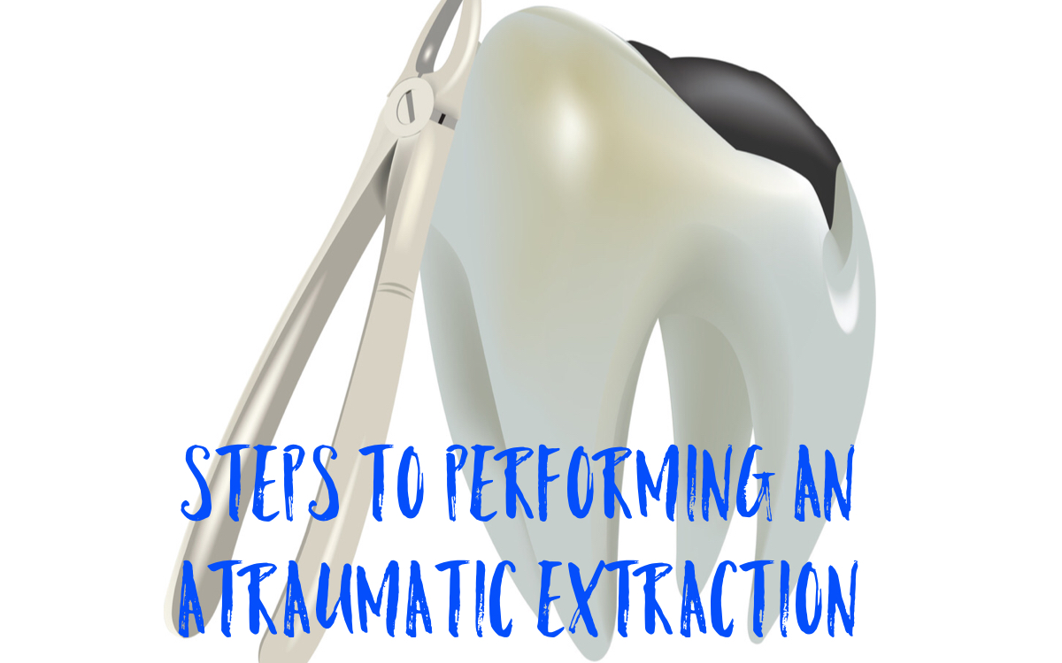
With the development of these new skills sets, a topic being thrown around most often these days is the concept of performing an atraumatic extraction, also referred to as, "performing a minimally invasive extraction", but what does that really mean? How does one effectively go about performing a minimally invasive extraction?
According to Dr. John Alonge, an accomplished oral surgeon who teaches minimally invasive extraction courses all over the world, "the concept of minimally invasive exodontia is based on preserving alveolar bone. Efficient and less traumatic exodontia should be part of a predictable routine for the general dentist and specialist." Dr Alonge often likes to remind his lecture participants how far extractions have come from what was originally taught in dental school. "Dental school teaches the conventional extraction techniques to either elevate the tooth by leveraging against the interproximal bone or use forceps to extract the tooth from its socket. The first technique frequently results in damage to the interproximal bone. The second technique often results in reshaping of the socket or alveolus." To avoid such issues, below are 3 easy steps to successfully achieve a minimally invasive extraction.
- Use of the Periotome or X-Otome to sever the PDL prior to performing an extraction. The Periotome has thin flexible blades that can be used by placing the instrument into the gingival sulcus and moved in an apical direction 360° around the tooth to sever the periodontal ligament (PDL). The X-Otome is quickly becoming more popular as it is more durable and easier to control compared with the periotome. The X-Otome is a hybrid, a cross between the periotome and a root elevator, that efficiently executes the severing of the PDL by rocking it slightly 2-3 degrees around the tooth structure. In addition to severing the periodontal fibers, the narrow tips preserve tissue structures and eliminate bone fracturing. The custom sure grip handle has a thumb rest for even power transfer of hand pressure. (Fig. 1)
- Use of the D-Lux elevators to apply slow and steady luxation forces around the crown or exposed root. Keep in mind the D Lux elevators are meant to be inserted vertically and used apically. It is advised to avoid the buccal/labial aspect of the tooth as this may result in unwanted alveolar bone fracture. (Fig. 2)
- Use of the X Trac or Plus Series forceps to apply slow and steady rotational forces clockwise and then counterclockwise until apical facial and lingual/palatal forces are applied. This rotation should be repeated several times until you feel slight mobility. Alonge suggests the tooth will generally release towards the facial and the sequence is the same regardless of whether the tooth is single or multi-rooted or the crown is missing or fractured. X-Trac and Plus Series forceps are instrumental when performing a minimally invasive extraction due to their unique beak designs that allow the forceps to slide further below the gum line to engage both the crown and the root at the same time. Additionally the serrations allow for greater grip and control when rotational forces are being applied. (Fig 3)
If you don't have the opportunity to sit in on one of Dr Alonge's hands on courses or lectures at an upcoming meeting, you can also access his Minimally Invasive Exodontia Techniques course through the continuing education portal of the ADA CE website. ACCESS NOW
Fig. 1 X-Otome in use Fig. 2 D-Lux Elevator in use Fig 3. 150+ Forceps in use